
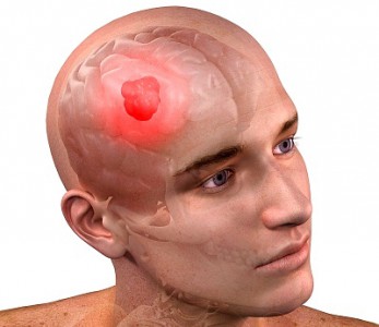
 Brain tumors are a heterogeneous group of various intracranial neoplasms, benign or malignant, that arise when a process of abnormal, uncontrolled division is triggered in cells that were once normal components of the brain tissue itself, the blood vessels of the brain, the cranial nerves, the meninges, the skull or the glandular structures of the brain, or that arise through metastasis of a primary tumor located in another organ.
Brain tumors are relatively rare – about 1.5% of all types of tumors. In Ukraine, the incidence was 4.6 per 100,000 population in 2006 and 4.4 per 100,000 in 2007.
Classification
Brain tumors can be divided into groups by the following features.
By primary site:
- Directly in the brain.
- Outside the brain (tumor metastasis).
By cell type:
- Neuroepithelial tumors (ependymoma, glioma, astrocytoma). They develop directly from brain tissue. They account for about 60%.
- Tumors of the meninges (meningioma). They develop from the tissues of the meninges.
- Pituitary tumors (pituitary adenoma). They form from cells of the pituitary gland.
- Cranial nerve tumors (neurinoma). They arise along the cranial nerves.
- Metastases from sites outside the brain. They reach the brain from other sites through metastasis.
- Dysembryogenetic tumors. They arise during embryonic development. A fairly rare but serious type of disease.
Distinctive features of brain tumors:
¨ Brain tumors very rarely spread beyond the CNS.
¨ Highly malignant tumors (meningioma, sarcoma, astrocytoma, medulloblastoma) can metastasize to the lungs in a small percentage of cases.
Medulloblastoma can metastasize to the lymph nodes, bones and bone marrow, and throughout the subarachnoid space of both the brain and the spinal cord.
Brain tumors grow in the enclosed space of the skull, and any additional growth compresses the brain, which makes the course of the disease more severe.
As they grow and enlarge, brain tumors block the flow of cerebrospinal fluid, causing blockage of the ventricles or occlusion of the subarachnoid space of the spinal cord, which results in hydrocephalus.
The location of the tumor inside the brain plays the main role in determining neurological symptoms and signs.
In some locations, brain tumors cause mental changes.
Clinical presentation
The clinical picture of brain tumors follows from their location in the limited volume of the cranial cavity. Compression or destruction of brain tissue in the area of the tumor (as the neoplasm grows into it) causes so-called primary, or focal, symptoms. As the disease progresses, so-called general cerebral symptoms appear, caused by impaired hemodynamics and intracranial hypertension.
Focal symptoms
Focal symptoms largely depend on the location of the tumor. The following groups can be distinguished:
- Sensory disturbances
- Motor disturbances (paresis, paralysis)
- Epileptic seizures
- Impaired hearing and speech recognition
- Impaired vision and recognition of objects and text
- Impaired spoken and written language
- Autonomic disorders
- Impaired coordination
- Psychomotor disorders
- Hallucinations
- General cerebral symptoms
General cerebral symptoms are symptoms that occur with increased intracranial pressure and compression of the main brain structures.
Headache:
In cancer, the headache is typically constant and intense, and it responds poorly to non-opioid painkillers. Lowering intracranial pressure brings relief.
Vomiting (unrelated to meals):
Vomiting of central origin usually occurs due to an effect on the vomiting center in the midbrain.
Dizziness:
It can result from compression of cerebellar structures. Dizziness can also be caused by tumor growth that impairs the blood supply to the brain.
Weakness, rapid fatigue:
This results from impaired blood supply to the brain (impaired outflow of blood or insufficient inflow).
Diagnosis
Because the tumor is located inside the skull, diagnosis is difficult. A cancer diagnosis is made definitively only after a histological report; without histology or cytology the diagnosis is not valid. The diagnosis of “brain tumor” is made in stages: first on an outpatient basis and then confirmed in the hospital. As a rule, the patient first sees a general practitioner (less often a neurologist or a doctor of another specialty). The patient seeks help because focal or general cerebral symptoms progress.
If the patient has neurological symptoms, he or she is referred to a neurologist for consultation.
The neurologist assesses the severity of the symptoms and makes a differential diagnosis. If a brain tumor is suspected, the patient is referred for computed tomography (CT) or magnetic resonance imaging (MRI). Contrast-enhanced MRI is the “gold standard” in diagnosing brain tumors. If a space-occupying lesion is found on the scan, a decision is made on admitting the patient to a specialized hospital.
Treatment
Treatment of brain tumors, like that of all cancers, is complex and quite expensive. All measures taken during the course of treatment can be divided into the following groups.
Symptomatic therapy
None of the measures in this group affect the cause of the disease itself; they only ease its course, helping to keep the patient alive or improve their quality of life.
Glucocorticosteroids (prednisolone) relieve swelling of the brain tissue and reduce general cerebral symptoms.
Antiemetics (metoclopramide) relieve vomiting caused by worsening general cerebral symptoms or following combined chemoradiotherapy.
Sedatives are used depending on the level of psychomotor agitation and the severity of mental disorders.
Nonsteroidal anti-inflammatory drugs (NSAIDs, for example, Ketonal) relieve pain.
Opioid analgesics (morphine, Omnopon) relieve pain, psychomotor agitation and vomiting of central origin.
Surgical treatment
Surgery is the main and most effective treatment for brain and spinal cord tumors, but removing a tumor from brain tissue is very difficult. Each such intervention is a complex neurosurgical operation. Since the surgeon has to remove the tumor with a margin of healthy tissue (to avoid metastasis), every such operation is traumatic, and quite often it is impossible altogether because the tumor is too large or is located in vital areas of the brain. Depending on the size, location and type of the tumor and the patient's condition, a decision is made on whether surgery is needed, how extensive it should be and how it should be performed. The use of modern laser and ultrasound technology in brain tumor surgery has made it somewhat more effective. The specific surgical approach and technique are chosen individually and depend on the tumor's location, size and cell type.
Radiation therapy
Radiation therapy plays an important role in treating brain tumors; its wide use and great importance are due to objective factors that limit surgery. When planning radiation therapy for patients with brain tumors, it is very important to determine an appropriate volume to be irradiated. For this purpose, data obtained during surgery and from preoperative and postoperative computed tomography, gammaencephalography and radioisotope studies should be used. Determining an appropriate radiation level makes it possible to avoid or minimize the negative effects of therapy. Radiation therapy works through the effect of ionizing radiation on the cells of tissues in the path of the radiation beam. Brain tumors are treated mainly with external beam gamma therapy.
Indications for radiation therapy:
All patients with malignant tumors after surgery.
Tumors located in areas where surgery is not possible because of the high risk associated with the tumor's anatomical location, as well as brainstem tumors.
Widespread tumors of both the brain and the spinal cord that make surgery pointless.
Regrowth of the tumor after treatment (at least 6 months later).
Contraindications to radiation therapy:
Benign tumors that are clearly demarcated and located in areas where they can be operated on.
Unclear location.
Unclear nature of the disease (a differential diagnosis is needed, for example with vascular changes or stroke).
A presumptive diagnosis of continued growth.
A high dose of previous irradiation and a short time since the first course.
Relative contraindications:
High intracranial pressure.
Low blood counts.
No morphological verification.
Brain structures differ in their radiosensitivity. The brainstem and the diencephalon (interbrain) are more sensitive to ionizing radiation – the total dose is 30–40 Gy. Cerebellar tissue has intermediate radiosensitivity – the total dose is 50–60 Gy. The structures of the cerebral hemispheres are more resistant to ionizing radiation. The total dose is 60–70 Gy.
Preoperative external beam gamma therapy is used very rarely. A postoperative course of external beam gamma therapy is used more often. Gamma therapy can also be given as a standalone treatment. There are several radiation therapy techniques:
Partial or local brain irradiation. It is used for small primary tumors, in the postoperative period once the tumor's location has been determined, and after whole-brain irradiation. The dose for local irradiation is about 60 Gy, with a single dose of 0.5–2.0 Gy. The aim of this technique is to spare the rest of the brain as much as possible. The irradiated volume includes the primary tumor with a 2 cm safety margin on all sides. CT data and the surgical report are used to determine the irradiated volume accurately.
Gamma therapy is given in courses in 2 stages, with irradiation 5 times a week and a two-day break. The first course lasts 21 days, followed by a three-week break.
Whole-brain irradiation is used for brain metastases and for palliative purposes. The irradiated volume includes the entire brain and the meninges. The total dose for whole-brain irradiation should not exceed 30 Gy; the single dose is 0.5–2 Gy, most often 1.8 Gy.
The optimal time to start radiation therapy is 14–21 days after surgery.
Radiation therapy is tailored individually to the tumor's cell type, size and location. Although it is effective, radiation therapy is quite hard on the patient and is accompanied by radiation reactions. Different radiation therapy regimens are designed for different types of tumors.
Chemotherapy
Chemotherapy drugs are used to treat some primary brain tumors, especially intermediate- and high-grade astrocytomas and medulloblastomas. The goal of drug treatment is to shrink the tumor and control its growth rather than to cure it. Chemotherapy is sometimes combined with radiation therapy, as this apparently improves the results of the latter. Hormonal drugs such as tamoxifen may also be given together with chemotherapy, which probably makes drug treatment more effective.
Follow-up care
After treatment is completed, the patient is discharged but remains registered for follow-up care. The patient is referred to the oncology center at their place of residence for repeat examinations. This system is aimed at the timely detection of tumor recurrence. After discharge, the patient should come three times at one-month intervals, then twice at three-month intervals, then twice at six-month intervals, and after that once a year. Follow-up care for people registered with an oncologist is lifelong.
Prognosis
The possibility of successful and complete treatment depends on timely and adequate diagnosis. With complete three-stage treatment started at an early stage of tumor development, five-year survival is 60–80%, depending on the histological type. If the patient seeks help late and surgery is not possible, five-year survival does not exceed 30–40%, depending on the histological type and the size of the tumor.
Prepared by: Tetiana Morhun, radiation oncologist, radiation therapy department No. 1.