
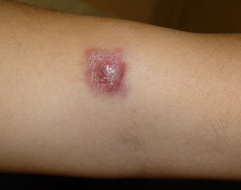
 Malignant cutaneous (skin) lymphoma is a systemic tumor process of lymphoid tissue with varying degrees of malignancy. Cutaneous lymphomas are a special case of lymphomas as a systemic tumor process in lymphoid tissue.
Prevalence. Malignant cutaneous lymphomas affect people of both sexes, more often men (male-to-female ratio 1.3:1). The disease can occur at any age, but is more common in middle and older age.
Risk factors Malignant cutaneous lymphoma is not a hereditary disease. Risk factors that increase the likelihood of developing it include occupational hazards in the chemical industry, construction and agriculture, increased radiation and sun exposure.
Causes
Malignant cutaneous lymphoma is based on neoplastic proliferation of clones of abnormal T lymphocytes, B lymphocytes and/or their precursors. Genetic factors are of great importance in the development of a malignant lymphoproliferative process in the skin.
Many researchers attribute the leading role in T-cell proliferation to retroviruses, primarily human T-lymphotropic virus type I (HTLV-1), as well as to various occupational hazards in the chemical industry, construction and agriculture. A link between cutaneous lymphomas and increased sun exposure has been noted.
Normal cells turn into tumor cells under the influence of the factors listed above and weakened immune defenses. Foci of lymphoid proliferation appear in the skin when a diagnosed or unrecognized nodal lymphoma spreads to it (secondary lymphoma), or when the skin is involved primarily at the early stages of the disease process (primary lymphomas).
Classification
Types include mycosis fungoides, poikilodermatous lymphoma, lymphosarcoma and others.
Mycosis fungoides Mycosis fungoides (fungoid mycosis) is the most clinically well-defined disease of this group and belongs to the T-cell lymphomas.
The classic form is divided into 3 clinical stages: nonspecific (eczematous-erythrodermic), infiltrative plaque and tumor. People of both sexes are affected; it occurs more often after the age of 30. It is accompanied by severe itching. Signs of all 3 stages can be seen on the skin of the same patient (clinical polymorphism).
In the nonspecific stage of mycosis fungoides the rash is nonspecific and may resemble eczema (more often seborrheic), pityriasis rosea, neurodermatitis, parapsoriasis en plaques or lichen planus. There is often marked palmoplantar hyperkeratosis with deep cracks. The rash responds poorly to treatment and recurs quickly. Patches become infiltrated and turn into plaques.
The time from the onset of the disease to the tumor stage of mycosis fungoides sometimes reaches decades. Plaques grow and turn into tumors; some tumors appear on unaffected skin and look like mushrooms. They are firm and elastic, pinkish-red in color and painless on palpation.
Gradually the tumors break down, and a crater-like ulcer appears.
In the tumor stage of mycosis fungoides the regional lymph nodes are affected; they are multiple, not fixed to the skin, and firm and elastic.
The patients' general condition and blood counts remain good right up to the terminal stage. Whether the disease can metastasize to internal organs remains an open question. Death results from cachexia, tumor breakdown, bleeding or secondary infection.
The erythrodermic form of mycosis fungoides spreads rapidly without a preceding rash. The entire skin looks red and swollen, with profuse scaling.
Poikilodermatous cutaneous lymphoma Poikilodermatous cutaneous lymphoma has a long, chronic, asymptomatic course and is resistant to treatment. Men are affected more often. Well-defined plaques of a red-brown or brownish color appear unnoticed, with fine scaling on the surface. Individual plaques can reach the size of a palm. There may be one or several of them.
Over a long time, a plaque becomes poikilodermatous: telangiectasias, atrophy, and hypo- and hyperpigmentation appear on its surface. The process changes little over time, but erythroderma (the erythrodermic form) may develop with its typical signs (chills, feeling unwell, fever; the lymph nodes are not enlarged when plaques are present). At the next stage, tumors appear on the plaques or outside them. This form of malignant cutaneous lymphoma is not accompanied by itching.
Lymphosarcoma Lymphosarcoma is more often a non-epidermotropic B-cell lymphoma. It is the fastest-progressing and most malignant of the malignant cutaneous lymphomas. There is no itching, and the rash is monomorphic. Without preceding stages, nodular indurations and tumors appear on normal skin. They are shiny and red with a livid (bluish) tint and are soft and elastic. Sometimes the rash takes the form of arcs, semi-arcs or discs. The lesions grow in size and number and quickly ulcerate.
Diagnosis
Differential diagnosis of malignant cutaneous lymphomas at the early stages includes common dermatoses (contact dermatitis, toxicoderma, pityriasis rosea, seborrheic eczema, Devergie's pityriasis rubra pilaris, parapsoriasis en plaques, lichen planus). The erythrodermic form is differentiated from psoriatic erythroderma, Devergie's pityriasis rubra pilaris and drug-induced skin lesions.
In the tumor stage, malignant cutaneous lymphoma is differentiated from various other neoplasms.
Laboratory diagnostic methods. Histological examination of the skin and lymph nodes, immunophenotyping, genotyping, polymerase chain reaction.
Treatment
Treatment of malignant cutaneous lymphoma depends on the grade of malignancy. At the early stages, local treatment may be enough – corticosteroid ointments and nitrogen mustard. At the initial stages of malignant proliferation with slow progression, retinoids and PUVA therapy are used. Electron beam therapy is used at all stages. For Sézary syndrome and erythrodermic variants of mycosis fungoides, leukapheresis and extracorporeal photopheresis are used.
At progressive stages, combination chemotherapy is given (cyclophosphamide, prospidine, doxorubicin, vincristine, prednisolone, etc.), more often with biological response modifiers – PUVA therapy, retinoids, cytokines and interferons (Reaferon, Gammaferon, Leukinferon).
Various combination chemotherapy regimens are used that achieve remission lasting from 1 to 10.5 months, depending on the type and stage of lymphoma, in 75% of cases. In severe cases, the CHOP (cyclophosphamide, doxorubicin, vincristine, prednisolone), COP (cyclophosphamide, vincristine, prednisolone) and CAMP (cyclophosphamide, methotrexate, 6-mercaptopurine, prednisolone, allopurinol) regimens are used. For Sézary syndrome, extracorporeal phototherapy (photopheresis) is effective. Tumors are treated with X-ray therapy.
Regimen and diet
No special regimen or diet is needed for malignant cutaneous lymphoma.
Prevention
Prevention of malignant cutaneous lymphoma means seeing a doctor early, at the first signs of the disease. Excessive sun exposure should be avoided.
Prognosis
After treatment with cytostatics and corticosteroids, immunity decreases, and pyoderma and generalized herpes may appear. The prognosis for recovery and life in malignant cutaneous lymphoma is unfavorable. Poikilodermatous lymphoma may follow a more benign course.
L. M. Bondarenko, head of the chemotherapy department, Sumy Regional Clinical Oncology Center