
 Preserving the nation’s reproductive health is an integral part of Ukraine’s demographic policy, so organizing measures aimed at improving the prevention, diagnosis and treatment of women with cancer of the reproductive organs remains a pressing problem that needs to be addressed at the national level.
Statistics show a steady rise in breast cancer incidence over the past 20 years, with no sign of stabilization. More than 1 million new cases of this disease are registered worldwide every year. In Ukraine, breast cancer incidence reached 67.1 cases per 100,000 women in 2012, 67.9 cases in 2013 and 58.7 in 2014. In the region, breast cancer incidence was 71.5 cases per 100,000 women in 2012, 67.8 cases in 2013 and 77.0 cases in 2014. While breast cancer incidence in Ukraine was 27.0 per 100,000 women in 1985, by 2013 it had risen to 67.9, meaning that it grew 2.5-fold over 30 years. In 2012, breast cancer ranked first among cancers in women in Ukraine, accounting for 19% of cases. More than 15,000 patients with breast cancer are diagnosed in our country every year, and about 7,500 women die of this cancer annually.
Introducing effective prevention and early diagnosis programs could be an effective way to improve the detection of malignant breast tumors. The breast cancer prevention and early diagnosis program has two main directions: primary prevention (etiopathogenetic) and secondary prevention, which consists of diagnosing and treating precancerous diseases.
Early diagnosis of breast cancer is possible if the following programs are carried out:
- Self-examination
- Preventive medical examinations, follow-up and treatment of pretumor conditions
- Screening examinations and mammography
It is known that in 80–90% of cases the disease is detected by the patients themselves, in 15–18% by a doctor during a preventive examination, and during mammography screening in 4–6% of those examined.
The breast is easily accessible for self-examination. You need to know how to do it. In Finland, girls are taught breast examination at school.
Breast self-examination (BSE) is a simple procedure that does not take much time and gives every woman a way to prolong her life. This simple procedure requires no special skills and can be included in your morning or evening routine. BSE helps a woman get to know her breasts and notice the slightest changes in their structure and function early.
The main rule of BSE is regularity. BSE should be done once a month, 7–10 days after the start of menstruation. If menstruation has already stopped, BSE is also done once a month (it is best to choose a fixed day, for example, the first day of each month).
If a woman finds a lump or other changes in her breasts, she should immediately see her local doctor or go to the outpatient clinic of the Sumy Regional Clinical Oncology Center. It should be noted that in nine cases out of ten the changes found are not malignant, but only a specialist can determine this. Self-examination cannot replace regular visits to the doctors of the women’s examination rooms that operate in the outpatient departments of all healthcare facilities.
Preventive medical examinations are based on clinical data (inspection, palpation) and data from additional examination methods: X-ray, morphological and ultrasound.
During the examination, attention should be paid to the symmetry, size and shape of the breasts and the condition of the nipple and areola, as well as to any abnormal masses and deformities, hyperemia, pigmentation and other skin changes. Superficial palpation is followed by deep bimanual palpation of the breasts. This provides information about the shape, size, consistency and surface of the tumor, its relation to the surrounding tissues and its mobility. The final stage of the physical examination is inspection and palpation of the axillary, subclavian and supraclavicular lymph nodes.
Unfortunately, no more than 15–17% of women undergo preventive examinations. This is especially true of certain age groups. The risk of breast cancer increases with age. While in women under 30 breast cancer occurs in 0.3–0.4% of cases, peak incidence is in the age group over 50 (70–80%).
Breast cancer screening is a method of detecting early forms of breast tumors through mass examinations of healthy women. Today, the standard for breast cancer screening programs is mammography and a clinical breast examination by a doctor. These methods complement each other. Such a program, called “Oncology”, is also in place in Ukraine. Thanks to the introduction of mammography screening programs in the economically developed countries of Europe and North America, there has been a significant increase in the diagnosis of breast cancer at the in situ stage, which accounts for 20–30% of cases.
Screening program
Breast cancer detection
| Age groups | Examination methods | Examination schedule |
| 20–35 years | Breast self-examination Examination by a doctor | Monthly Twice a year |
| 35–40 years | Breast self-examination Examination by a doctor Baseline mammography | Monthly Twice a year as a reference for comparison |
| 40–50 years | Breast self-examination Examination by a doctor Mammography combined with ultrasound | Monthly Twice a year Every 1–2 years |
| Over 50 years | Breast self-examination Examination by a doctor Mammography combined with ultrasound | Monthly Twice a year Annually |
X-ray examination is one of the main methods for diagnosing various forms of breast cancer.
Non-contrast mammography is the most developed and widely used additional method of breast examination. It is informative enough not only to identify an abnormal lesion and make a diagnosis, but also to assess the growth pattern and spread of the tumor, which is extremely important for choosing a treatment strategy. Mammography can detect lumps in breast tissue as small as 0.3 cm, as well as the tiniest calcifications, which are direct signs of breast cancer. That is why mammography should be widely used in preventive examinations of women at increased risk of breast cancer.
Thermography is a highly sensitive method of recording infrared radiation that can detect temperature differences of 0.1–0.01 °C between symmetrical areas of the breasts. In malignant tumors, the temperature difference between the healthy and the affected breast reaches 1.5–2 °C, which makes it possible to use this method to diagnose early forms of cancer during preventive examinations of women.
Ultrasound diagnostics of breast tumors is based on the difference in the echographic image of examined tissues of different density. The information obtained by ultrasound makes it possible to assess the tumor process, its location, shape and size objectively.
Radioisotope examination is based on the increased uptake of certain substances (phosphorus, gallium citrate, technetium, etc.) by malignant cells compared with normal cells. Some time after these radioactive agents are introduced into the body, a counter is used to measure how much of them has accumulated in the breasts. This method has proved useful for diagnosing breast cancer metastases in the bones, liver and lymph nodes.
Morphological confirmation of the clinical diagnosis is obtained by needle biopsy of the tumor or a lymph node, as well as by cytological examination of nipple discharge. In some cases, an excisional biopsy of the breast tumor with rapid intraoperative diagnosis is performed.
Breast cancer treatment methods
- Surgery
- Polychemotherapy (systemic and regional)
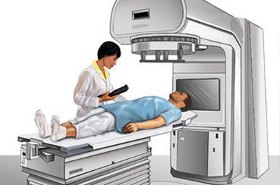
- Radiation therapy
- Hormone therapy
Treatment of breast cancer is individualized and depends primarily on the stage of the disease, the growth pattern of the tumor, its morphological features (growth rate, grade of malignancy), the patient’s age, the state of her reproductive function and any accompanying diseases. Treatment includes a number of simultaneous and sequential measures and is therefore, as a rule, combined and multimodal. In early preclinical stages and at stage T1-2N0M0, with nodular forms and an outer (lateral) location of the tumor, organ-preserving surgery is acceptable in the form of a radical resection of the breast or a quadrantectomy followed by irradiation of the remaining part of the breast. For medial and central cancers of stages I–IIa, as well as stage IIb (T2 N1), treatment begins with preoperative external beam gamma therapy and polychemotherapy followed by surgery. In the postoperative period, the parasternal and subclavian areas are also irradiated, and adjuvant courses of polychemotherapy are prescribed.
In stages III and IV of breast cancer (locally advanced disease), preoperative polychemotherapy (2–4 cycles) is given, followed by radical mastectomy with pre- and postoperative external beam gamma therapy to both the primary tumor and all areas of regional metastasis. In the six months after surgery, 4–6 cycles of polychemotherapy and hormone therapy (antiestrogens, aromatase inhibitors) are prescribed.
In advanced cases and in certain clinical forms of breast cancer with an aggressive clinical course (mastitis-like, erysipelas-like, edematous, en cuirasse), conservative treatment is indicated – radiation therapy and chemohormonal therapy.
Hormonal treatment of cancer at this site is based on the hormone dependence of the breast. Suppressing or switching off the function of the organs that stimulate the proliferation of the glandular epithelium of the breast leads to atrophy of the gland structures and, at the same time, to regression of the cancer.
Production of the relevant hormones is switched off surgically, with radiation or with medication. For this purpose, surgical oophorectomy is used. In recent years, luteinizing hormone-releasing hormone analogs (goserelin – Zoladex, buserelin – Suprefact, etc.) have been used increasingly often. Hormone therapy works in about one third of breast cancer patients. To predict the clinical effect of hormone therapy, the patient’s hormonal status and the presence of estrogen and progesterone receptors on the surface of the cancer cells are taken into account. Experience has shown that patients whose tumors lack hormone receptors do not respond to hormone therapy.
Androgens are used when menstrual function is preserved. They are useful for bone metastases. Estrogens are used in patients in deep menopause. The effect of androgens and estrogens is explained by suppression of pituitary function.
Corticosteroids also suppress the function of the adrenal cortex and thus reduce estrogen secretion. That is why corticosteroids are especially indicated for patients in menopause.
The hormone therapy of choice for patients of menopausal age is antiestrogens and/or aromatase inhibitors (provided the tumor has estrogen receptors).
Prognosis. The effectiveness of therapy depends on the stage of the disease. After combined treatment, the overall 5-year survival rate is
50–60%.
V. I. Konanykhin, Chief Physician, Sumy Regional Clinical Oncology Center